Tag Archives: Brain Science

Self Organized Criticality and Brains

Waking Brain

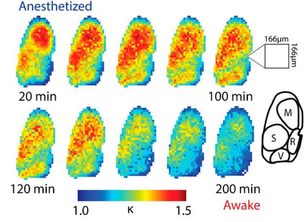
Figure 1. The above series of images show the deeply anesthetized brain (top left) at 20 minute intervals (from left to right) until the brain is in the fully awake and alert state (bottom right). All regions tended towards k ≈ 1 (blue), or criticality, as mice recovered from anesthesia toward the resting state. k < 1 is subcritical (not shown in figure) and k > 1 is supercritical. From Figure 8a in the paper “Voltage Imaging of Waking Mouse Cortex Reveals Emergence of Critical Neuronal Dynamics” published December 10, 2014 in The Journal of Neuroscience.

Criticality predicts a power law relationship in the brain. It also predicts that cortical dynamics should be independent of scale. In my previous post Engineered Proteins Enable Watching High Resolution Electrical Activity Across the Brain we saw that a recently published study (“Voltage Imaging of Waking Mouse Cortex Reveals Emergence of Critical Neuronal Dynamics” published December 10, 2014 in The Journal of Neuroscience) measured a power law relationship in awake brains consistent with brain dynamics adhering to the physical laws of self organizing criticality.

Are cortical dynamics independent of scale? The team looked to see if the observed cascade size distributions maintain the same form independent of scale, location, and resolution in a resting awake, but not in an anesthetized, animal. They calculated k values within small subregions of about two-tenths of a percent of the total cortical region imaged. These values were highly variable across the anesthetized cortex but for each subregion they converged towards k = 1 as the animal transitioned into the awake and resting state (see Figure 1 above).

In addition, k values were calculated across cortical subregions at 3 levels of spatial averaging across the raw data images. A raw data image was comprised of 15,676 pixels (each pixel averaged the signal from 33 square micrometers of cortex). Fine level images contained 3971 pixels (spatial average across 4 pixels), medium contained 290 pixels (spatial average across 54 pixels), and course contained 21 pixels (spatial average across 747 pixels). In the anesthetized state k values changed with scale (resolution) but in the awake and resting animal k values remained close to 1 at every scale. The research team concluded that cortical dynamics are independent of scale in the awake and resting brain but are scale dependent in the anesthetized animal.

Does the idea of criticality and its potential action in the brain provide us with useful insight to help us understand how our brains work? Neuroscience continues to lack a general theory describing small circuit and large network signal processing in the brain. To this end, a mathematical law or set of laws that may capture important features of observed brain dynamics is an exciting contribution.

The idea of self organized critical states derives from statistical mechanics. The behavior of complex systems can be organized in different types or phases separated one from the other by a sharp boundary or phase transition. There is a lot of very interesting work describing the electrical activity of individual neurons in terms of phases and phase transitions (see for example Dynamical Systems and Silicon Based Hybrid Spiking Neurons). When the system transitions from one phase to another, it experiences dramatic changes in behavior. At certain (second-order) phase transitions, fluctuations occur at all length scales and the system exhibits scale invariance and power-law behavior. Self organized critical systems are defined by second-order phase transitions.

Current results suggest that cerebral cortices function as self organized critical systems while awake and at rest. Cortex under pentobarbital anesthesia functions in the supercritical (k > 1) range of phases according to these data. It’ll be interesting to see how well this organizing principle holds across the many organs of the brain.

Self Organized Critical States and Our Brains

SOCSandPile

Figure 1. The sandpile model of self-organized criticality from Figure 1 in the book “How Nature Works: the science of self-organized criticality” published 1996.

“…consider the scenario of a child at the beach letting sand trickle down to form a pile (Figure 1). In the beginning, the pile is flat, and the individual grains remain close to where they land. Their motion can be understood in terms of their physical properties. As the process continues, the pile becomes steeper, and there will be little sand slides. As time goes on, the sand slides become bigger and bigger. Eventually, some of the sand slides may even span all or most of the pile. At that point, the system is far out of balance, and its behavior can no longer be understood in terms of the behavior of the individual grains. The avalanches form a dynamic of their own, which can be understood only from a holistic description of the properties of the entire pile rather than from a reductionist description of individual grains: the sandpile is a complex system.”

Per Bak, “How Nature Works: the science of self-organized criticality” (1996)

Living organisms operate at stable points far from equilibrium. Homeostasis, a process characteristic of living things, maintains the set point (or points) far from equilibrium. Even the simplest of life forms such as viruses exist far from equilibrium. The genetic code they carry is information that takes energy to get in formation and to maintain. Equilibrium, lack of information, and death seem synonymous. So it probably isn’t a surprise when we say that brain activity is a process that is carried out in states far from equilibrium.

That said, non-living things can exist at stable points far from equilibrium. The whole idea first came to my attention with the work of Ilya Prigogine, the 1977 Nobel Prize winner in Chemistry. Prigogine mixed chemicals together that, when energy was added to the mixture, self organized into intricate structures. These structures sucked in and used energy. He called them dissipative structures. Dissipative structures are self organized systems that exist at set points far from equilibrium. If energy input is cut off, the dissipative structure falls back into chaos and then randomness. To equilibrium.

Around 1987 the physicist Per Bak introduced the idea of criticality. Per Bak pointed out that a large amount of variability in our universe is the normal rather than exceptional condition and he defined systems with large variability as complex. He said these complex systems that exist far from equilibrium exhibit universal phenomena no matter if they are weather patterns, a biological organism, or a chemical reaction.

In my previous post Engineered Proteins Enable Watching High Resolution Electrical Activity Across the Brain, the research team that published the paper “Voltage Imaging of Waking Mouse Cortex Reveals Emergence of Critical Neuronal Dynamics” published December 10, 2014 in The Journal of Neuroscience was particularly interested in finding out if activity in the mammalian cortex adhere to the rules of critical dynamics.

Cascade size probability distributions approach power-law form during recovery from anesthesia

Figure 2. Cascade size probability distributions approach power-law form during recovery from anesthesia. Figure 3 in the paper “Voltage Imaging of Waking Mouse Cortex Reveals Emergence of Critical Neuronal Dynamics” (published December 10, 2014 in The Journal of Neuroscience)

While recording electrical activity across mouse brains, the research team saw cascades of various sizes travel across the cerebral cortex (for a movie and more information see Engineered Proteins Enable Watching High Resolution Electrical Activity Across the Brain). Locally isolated and short-lived electrical cascades were observed more often than large cascades. In the anesthetized state, very large cascades of activity were seen that were not usually seen in other brain states.

The probability of observing a cascade of a particular size was used as a measure of cortical dynamics during discrete 20 minute time periods (see Figure 2 above). For example, each blue point in the Figure 2A left hand graph is the probability that a cascade of the size displayed on the x-axis (notice the x-axis is in logarithmic scale) occurs in the anesthetized mouse’s cortex. The blue points correspond to the time period represented by the blue bar (the first 20 minutes of the experiment) in the bar chart in Figure 2C. In the same way, each red point in the Figure 2A right hand graph represents the probability that a cascade of the size displayed on the x-axis occurs in the awake mouse’s cortex. The awake time period is represented by the red bar (the last 20 minutes of the experiment from 180 to 200 minutes) in the bar chart in Figure 2C.

These data show that the probability of observing a cascade of a particular size in the anesthetized mouse does not follow a power law relationship but in the awake animal it does. The lack of a power law relationship is consistent with the lack of criticality in an anesthetized brain and the the power law relationship measured in awake brains is consistent with brain dynamics adhering to the physical laws of self organizing criticality.

Criticality predicts a power law relationship in the brain. It also predicts that cortical dynamics should be independent of scale. In my next post we’ll look at how this research team addresses the question of scale independence. We’ll also consider how this work may provide us with useful insight to help us learn more about how our brains work.

Engineered Proteins Enable Watching High Resolution Electrical Activity Across the Brain

Movie 1. Video of voltage cascades across the mouse brain from Movie 1 in the paper “Voltage Imaging of Waking Mouse Cortex Reveals Emergence of Critical Neuronal Dynamics” published December 10, 2014 in The Journal of Neuroscience.

Genetically encoded voltage indicator proteins have the potential to move the electrophysiological investigation of brain function into a new era by enabling both small scale and large scale investigation of electrical signaling in brains. Research reported recently in the paper “Voltage Imaging of Waking Mouse Cortex Reveals Emergence of Critical Neuronal Dynamics” (published December 10, 2014 in The Journal of Neuroscience) used optical imaging of a genetically encoded voltage indicator expressed in layer 2/3 neurons of the mouse cortex to investigate the relationship between small scale and large scale signal processing in the cerebral cortex.

Microelectrode recordings usually focus on the activity of a single cell, tens of microns in size, and provide fine temporal resolution down to the sub-microsecond level. In contrast, electroencephalography (EEG) records data from across large expanses of cortical tissue with high temporal resolution but poor spatial resolution, making it difficult to impossible to discern small scale signal processing characteristics. Hi spatial resolution across an entire brain may be achieved using functional Magnetic Resonance Imaging (fMRI) but with a severe loss of temporal resolution. The technique used in the study under review, optical imaging of a genetically encoded voltage indicator expressed in layer 2/3 neurons, enables capturing electrical activity across an entire hemisphere of the mouse cerebral cortex down to a 33 x 33 micrometer individual pixel resolution at a temporal resolution down to 20 milliseconds.

It’s unclear how results using this new technique will map to results gathered using older methods. In particular, how is a single neuron response represented in the data gathered at these resolutions? Does it matter or is the mass action of neurons more relevant at the recorded spatiotemporal resolution? Whatever the answers to these questions may be, this new technique has provided the research team with the ability to measure electrical signal processing in layer 2/3 cortex at a relatively small scale and then to use averaging techniques to look at the same signals at much lower resolutions. In this way, they could investigate affects of scale on signal processing in the cerebral cortex.

The signal processing paradigm used in this experiment was very simple. Measure cortical responses from mice beginning while they were under pentobarbital anesthesia, through their emergence from the anesthetic, until they were awake and at rest. Under all states, the research team saw cascades of activity sweep across the cerebral cortex (see Movie 1 above). However, the nature of the cascades depended on the state of the animal.

Across all brain states the researchers saw small, locally isolated and short-lived cascades more often than large cascades. In the anesthetized state, however, very large cascades of activity were often seen that were not usually seen in other brain states. Regional variations in cascade statistics (the probability of observing a cascade of a particular size) under anesthesia displayed clear anatomical boundaries associated with known sensory and motor regions. These boundaries disappeared during recovery phase until you’d be hard pressed to see clear anatomical distinctions in the cascade statistics in the awake animal.

The authors of this paper were looking for evidence to help them answer two questions: 1) Are cortical dynamics the same or different at different scales of extent? Or are cortical dynamics independent of scale? and 2) Does activity in the mammalian cortex adhere to the rules of critical dynamics?

I will address these questions in light of their findings in my next post.